Instrumentation
The CSCI Flow Cytometry core maintains an array of high-end, cutting-edge, and well-maintained instruments to serve the diverse range of flow cytometry and cell sorting needs across Columbia University.
| Category | Instrument | Location |
|---|---|---|
| Conventional Cell Sorting | Sony MA900 (2) | VP&S 11511 |
| Waters Biosciences FACSAria (1) | VP&S 11511 | |
| Spectral Cell Sorting | Thermo Fisher Invitrogen Bigfoot (1) | VP&S 11511 |
| Image-Enabled Spectral Cell Sorting | Waters Biosciences FACSDiscover S8 (2) | VP&S 11511 and Hammer 1211A |
| Conventional Cell Analysis | Agilent NovoCyte Penteon (3) | VP&S 11501 and Hammer 1211A |
| Agilent NovoCyte Quanteon (1) | VP&S 11501 | |
| Spectral Cell Analysis | Sony ID7000 Spectral Cell Analyzer (1) | VP&S 11501 |
| Imaging Flow Cytometry | Cytek Amins ImageStream-X MkII | VP&S 11501 |
| Single Cell Capture for Genomics | 10x Genomics Chromium X | BB 1116 |
| Waters Biosciences Rhapsody Scanner and HT | BB 1116 | |
| qPCR | Bio-Rad CFX Opus 96 and 384 | BB 1119 |
| Advanced Imaging |
EVOS S1000 Spatial Imaging System Managed by the Stem Cell Core |
BB1120 |
| Basic Imaging | Thermo Fisher EVOS FLoid Imager | VP&S 11511 |
| Cell Counting |
Nexcelom Cellometer Auto 2000 Cell Viability Counter |
BB 1116 |
Conventional Cell Sorting
Sony MA900 Multi Application Cell Sorter
OVERVIEW

CSCI Flow Cytometry maintains two Sony MA900 cell sorters: "Mars" and "Venus".
The Sony MA900 is a benchtop cell sorter equipped with 4 lasers (405 nm, 488nm, 561 nm, 638 nm) and 14 optical parameters (12 fluorescence channels and 2 light scatter channels). The MA900 is built around its highly innovative and proprietary sorting chip technology, which combines microfluidics with traditional electrostatic droplet sorting.
The MA900 can simultaneously collect up to 4 unique cell populations into tubes (1.5, 5, and 15 mL) and is capable of direct deposition down to as low as a single cell into multi-well plates.
A notable aspect of the Sony MA900 is its extraordinary ease of use and automation capabilities, and new users can learn to operate the Sony MA900 in as little as 3 hours.
OPTICAL CONFIGURATION OVERVIEW
4 lasers + 14 PMT detectors (12 fluorescence + 2 light scatter)
- 488 nm and 561 nm lasers (collinear): 5 PMT detectors
- 405 nm and 638 nm lasers (collinear): 7 PMT detectors
- Light scatter parameters: 488 nm forward scatter (FSC) and back scatter (BSC)
FEATURES AND CAPABILITIES
- Chips are available in multiple nozzle sizes accommodate a wide variety of cell types: 70 μm (40 PSI), 100 μm (20 PSI), 130 μm (9 PSI).
- Multiway sorting (up to 4-way) into 1.5 mL, 5 mL, and 15 mL tubes
- Direct plate deposition, including single cell sorting, into a variety of plate types
- Temperature control of both sample input and output
- Demonstrated high performance in purity, recovery and viability
- Housed in a Class II biosafety cabinet for sorting of primary human tissue
Waters Biosciences FACSAria SORP Cell Sorter
OVERVIEW
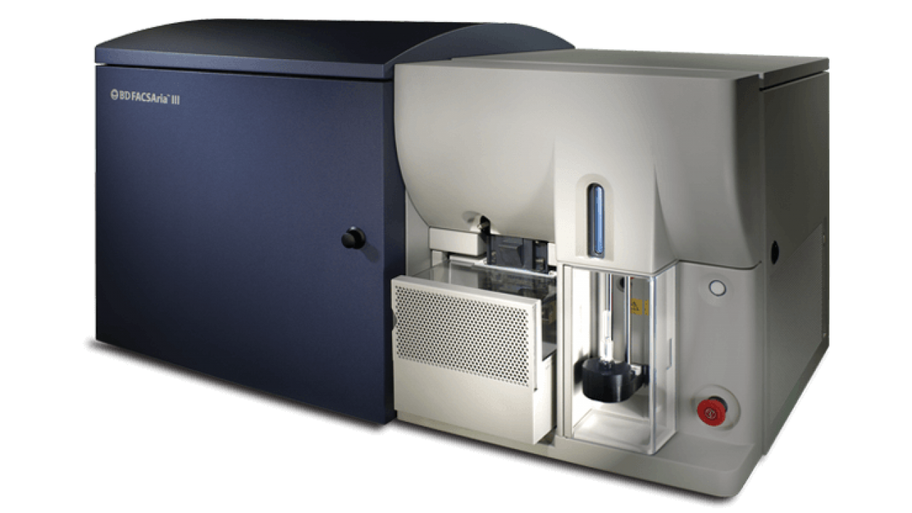
CSCI Flow Cytometry maintains one 20-parameter, 18-color cell Wates Biosciences (former BD Biosciences) FACSAria cell sorter, FACSAria “Neptune”, equipped with 5 high-powered, spatially-separated laser lines. The FACSAria is a workhorse for consistent isolation of cells with high purity, recovery and viability.
OPTICAL CONFIGURATION OVERVIEW
5 lasers + 20 optical detectors (18 fluorescence + 2 light scatter)
- 355 nm (60 mW): 3 PMT detectors
- 405 nm (100 mW): 6 PMT detectors
- 488 nm (100 mW): 3 PMT detectors
- 561 nm (100 nW): 4 PMT detectors
- 637 nm (140 mW): 3 PMT detectors
- Light scatter: 488 nm forward scatter (photodiode) + SSC
FEATURES AND CAPABILITIES
- Highly stable fixed-alignment optics which greatly simplify setup and troubleshooting
- Next-generation square cuvette which offers superior fluorescence sensitivity and superb recovery performance for cells of all sizes, including large and fragile cells
- Three nozzle sizes - 70 μm (70 PSI), 100 μm (20 PSI), and 130 μm (12 PSI) to accommodate cell types of various sizes and robustness
- 4-way sorting into a variety of tube types, including 1.5 mL, 5 mL, and 15 mL tubes (2-way sorting only into 15 mL tubes)
- Direct deposition into a variety of plate types, including 96-well, 384-well, and Terasaki plates
- Temperature control of both sample input and sorted fraction
- Integrated bubble detector on sample path, allowing sorting of entire volume sample without the worry of introduction of air bubbles into the sample fluidics
- Aerosol Management Systems (AMS) for biosafety concerns
- Housed in a Class I biosafety cabinet (BioBubble) for sorting of primary human tissue
Spectral Cell Sorting
Thermo Fisher Invitrogen Bigfoot Spectral Cell Sorter
OVERVIEW

The CSCI Flow Cytometry Core maintains one Thermo Fisher Invitrogen Bigfoot Spectral Cell Sorter.
The Thermo Fisher Invitrogen Bigfoot is a state-of-the-art cell sorter equipped with a variety of innovative and unique features not found on any other instrument and has demonstrated high-performance and versatility for a wide range of cell sorting experiments.
A notable aspect of the Bigfoot is its speed, and it is the fastest cell sorter maintained by the core facility. Built on sense-in-air fluidics and the narrow pulse width of signals generated on these types of systems, the Bigfoot can process data at extremely high rates with no dead time and thus no hard/electronic aborts. In addition, the Bigfoot’s flow rate is significantly faster than other cell sorters, and sample can be acquired at up to approximately 120 µl/min, even for sorting.
Importantly, the Bigfoot's speed does not come at the expense of cell viability, recovery, or purity — metrics on which the instrument consistently demonstrates outstanding performance.
OPTICAL CONFIGURATION OVERVIEW
5 lasers + 53 optical detectors (48 fluorescence + 5 light scatter)
- 349 nm (100 mW): 12 PMT detectors
- 405 nm (100 mW): 13 PMT detectors (12 fluorescence + 1 light scatter)
- 488 nm (125 mW): 11 PMT detectors (7 fluorescence + 4 light scatter)
- 561 nm (100 mW): 12 PMT detectors
- 640 nm (100 mW): 5 PMT detectors
- Light scatter: 488 nm forward scatter and side scatter, 488 nm depolarized FSC and SSC, 405 FSC (small particle detector)
FEATURES AND CAPABILITIES:
- Choose between spectral unmixing and conventional (compensation) workflows for fluorescence detection.
- Multiple nozzles sizes to accommodate a variety of cell types: 70 µm (60 PSI), 85 µm (40 PSI) 100 µm (30 PSI), 120 µm (20 PSI), 150 µm (12 PSI) and 200 µm (6 PSI)
- Multi-tube loader accommodates up to 6 samples with integrated temperature control and agitation. Compatible tube types are 1.5 mL, 5 mL, and 15 mL
- High-speed data processing capabilities facilitate ultra-fast sorting
- Fluidics containers that are swappable without turning off the stream
- Full software automation of setup and maintenance tasks
- Startup – one-button startup can be schedule ahead of time
- Nozzle alignment, QC, and droplet/stream setup
- Nozzle unclog
- Cleaning
- Shutdown
- Decontamination
- Integrated biosafety cabinet for Class II protection
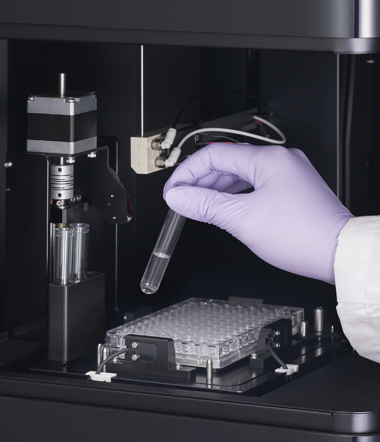
Image-Enabled and Spectral Sorting
Funded by the National Institutes of Health under award number S10OD036289 (BD FACSDiscover S8 "Odyssey")
Waters Biosciences FACSDiscover S8

Overview
The CSCI Flow Cytometry Core mantains two BD FACSDiscover Cell Sorters, "Odyssey" and "Loki".
The Waters Biosciences FACSDiscover S8 Image-Enabled Spectral Cell Sorter is the first instrument that is capable of cell sorting based on imaging data. Patented CellView™️ high-speed imaging technology facilitates spatially resolved signal measurements without a camera by using the optical and electronic components available on flow cytometers.
In addition to image-enabled sorting, the FACSDiscover S8 incorporates a 5-laser, 78 detector spectral detection system for true high parameter sorting.
OPTICAL CONFIGURATION OVERVIEW
5 lasers + 86 total detectors
Imaging Configuration:
(All imaging detectors are photomultiplier tubes)
- 488 nm laser (100 nm): 3 PMT fluorescence detectors + 3 PMT light scatter detectors
Spectral Configuration:
(All spectral detectors are avalanche photodiodes)
- 349 nm (30 mW): 22 ADP (avalanche photodiode) detectors
- 405 nm (50 mW): 20 ADP detectors + 2 PMT light scatter detectors
- 488 nm (100 mW): 16 ADP detectors
- 561 nm (50 mW): 12 ADP detectors
- 640 nm (100 mW): 8 ADP detectors
Light Scatter Detectors (photomultiplier tubes):
- Imaging Light Scatter: 488 nm forward scatter + side scatter + axial light loss (ALL)
- Traditional light scatter: 405 nm axial light loss (ALL) + 405 nm side scatter
FEATURES AND CAPABILITIES
Image-Enabled Sorting: Utilize quantified imaging data (“features”) in your sort logic. Imaging features can be plotted and gated like traditional flow cytometry data and used for sorting. Below are some examples of what image-enabled sorting enables:
- Differentiation of punctate from diffuse fluorescence signal
- Detection of the degree of colocalization of two fluorescence signals
- Measurement of the distance between two fluorescence signals
- Detection of cell-cell interactions
- Enhanced doublet exclusion
- Sorting cells based on cell size
Imaging Configuration in depth:
- Six Imaging Channels (all based on 488 nm laser illumination)
- 3 morphological/light scatter images: forward scatter (FSC), side scatter (SSC), and axial light loss (ALL)
- 3 fluorescence images:
- FL1: LP505, 534/46 (FITC, AF488, GFP, YFP, etc)
- FL2: LP570, 600/60 (PE, tdTomato, RFP, mScarlet, RY586, etc.)
- FL3: LP675, 788/225 (DRAQ5, PE-Cy7, RB705, RB780)
- Magnification: ~15x
- Resolution: 1.2 μm^2/pixel
- Field of View: 60 μm
Imaging Features:
Imaging features permit quantification of imaging measurements for biological measurements. These quantified features can be plotted like flow cytometry data and used for sorting. The following features are available on the FACSDiscover S8: Light Loss, Forward Scatter, Side Scatter, Correlation, Delta Center of Mass, Diffusivity, Eccentricity, Max Intensity, Radial Moment, Center of Mass (X), Center of Mass (Y), Long Moment, Short Moment, Size, Total Intensity.
Spectral Cell Sorting. The FACSDiscover S8 is equipped with a state-of-the-art spectral detection system coupled with BD patented SpectralFX™️ electronics and algorithms.
Highlights of SpectralFX™ include:
- Full spectrum optics and optimized hardware design with 78 solid state detectors and 5 lasers for all the classical benefits of spectral flow cytometry.
- System aware unmixing algorithm adapts to your sample and instrument in real time to manage spread.
- Next generation QC system uses LEDs and beads to measure noise, perform gain calibration and provide real time noise/signal hardware information.
- Guided workflow allows users to learn quickly and use best practices in experimental setup.
Spectral Configuration:
The FACSDiscover S8’s SpectralFX™ technology is equipped with the following hardware components for spectral detection.
- Five spatially separated excitation lasers
- 349 nm (UV), 30 mW
- 405 nm (violet), 50 mW
- 488 nm (blue), 100 mW (488 nm laser is split between the SpectralFX and CellView systems)
- 561 nm (yellow green), 50 mW
- 637 nm (red), 100 mW
- 78 fluorescence avalanche photodiode (ADP) photodetectors
- 349 nm: 22 detectors, 365 - 860 nm
- 405 nm: 20 detectors, 410 - 860 nm
- 488 nm: 16 detectors, 495 - 860 nm
- 561 nm: 12 detectors, 570 - 860 nm
- 637 nm: 8 detectors, 645 - 860 nm
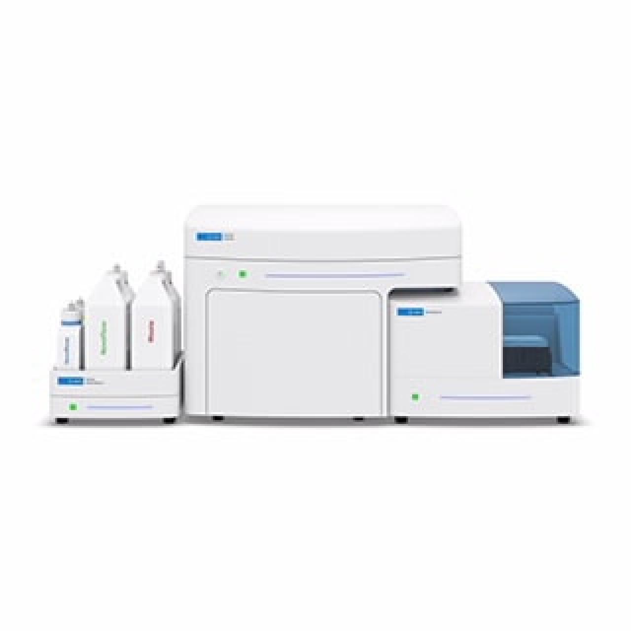
Spectral Flow Cytometry
Sony ID7000 Spectral Cell Analyzer

OVERVIEW
The CSCI Flow Cytometry Core maintains one Sony ID7000 Spectral Cell Analyzer, "Sirius".
The Sony ID7000 Spectral Cell Analzyer is a cutting-edge spectral flow cytometer analyzer equipped with 5 lasers and 147 fluorescence photomultiplier tube (PMT) detectors giving the instrument the ability to detect more than 40 fluorochromes in a single sample. The full spectrum detection capabilities of the instrument permit measurements of virtually any excitable fluorochrome without dedicated filter sets. In addition, by measuring spectral signatures of each fluourochrome, the ID7000 can detect the presence of nearly overlapping dyes. Furthermore, autofluorescence can be treated as a unique cellular parameter and subtracted from total fluorescence, reducing background and increasing resolution.
At the heart of the ID7000 is the best-in-class auto-loader capable of true hands-free sample
acquisition from a variety of plate types, including both 96 and 384-well formats, and a multi-tube rack. Temperature control and sample mixing are integrated into the autoloader and can be programmed and tailored uniquely to each experiment. Intuitive ID7000 Software simplifies the workflow for setting up a high parameter experiment, collecting data, and accurately performing spectral unmixing.
OPTICAL CONFIGURATION OVERVIEW
5 lasers + 149 detectors (147 fluorescence + 2 light scatter)
Full-spectrum detection from 361 nm – 844 nm
- 355 nm (50 mW): 35 independent detectors (3 single-channel PMTs + one 32 channel PMT)
- 405 nm (100 mW): 35 independent detectors (3 single-channel PMTs + one 32 channel PMT)
- 488 nm: 32 independent detectors (32-channel PMT)
- 561 nm: 26 independent detectors (multichannel PMT
- 637 nm637 nm laser 19 independent detectors (multichannel PMT)
- Light scatter: 488 nm forward scatter (FSC) and side scatter (SSC)
FEATURES AND CAPABILITIES
- Full spectrum detection from 361 nm – 844 nm across 5 lasers
- Advanced spectral unmixing capabilities using the Weighted Least Squares Method (WLSM), which facilitates highly accurate unmixing.
- Standardization Mode: instrument settings are standardized based on daily QC results for highly consistent results between ID7000 instruments and from day to day. Standardization Mode also allows you to record experimental samples at any PMT voltage without having to rerun single color controls.
- Spectral reference library allows you save spectral signatures to a catalog to use across experiments and user accounts.
- Ability to use compensation beads for virtually any fluorochrome
- Walk-away operation with the self-calibrating autoloader. Autoloader also integrates temperature control and sample mixing.
- Automatic clog monitoring
Conventional Flow Cytometry
Agilent NovoCyte Penteon and Quanteon
OVERVIEW
The CSCI Flow Cytometry Core maintains 4 Agilent NovoCyte cell analyzers, NovoCyte Penteon "Athena", "Saturn", and "Freya", and NovoCyte Quanteon "Diana".
The Agilent NovoCyte cell analyzer platform is a flexible reliable conventional flow cytometer workhorse analyzer suitable for a variety of applications. Controlled by extremely well-designed and powerful NovoExpress software, the NovoCyte is a robust, easy-to-use analyzer platform capable of handling a range of experiments, including those requiring measurement of at least 20 colors. Equipped with high-powered lasers and unique silicon photomultiplier (SiPM) detectors that require only minimal gain adjustments and facilitate a 7.2-decade log scale, the NovoCyte is capable of sensitive and consistent measurements.
OPTICAL CONFIGURATION OVERVIEW
NovoCyte detectors are silicon photomultipliers (SiPMs)
Penteon:
5 lasers + 32 optical detectors (30 fluorescence + 2 light scatter)
- 349 nm (20 mW): 7 SiPM detectors
- 405 nm (100 mW): 7 SiPM detectors
- 488 nm (100 mW): 8 SiPM detectors (7 fluorescence + 2 light scatter)
- 561 nm (100 mW): 6 SiPM detectors
- 640 nm (100 mW): 4 SiPM detectors
- Light scatter: 488 nm forward scatter (FSC) and side scatter (SSC)
Quanteon:
4 lasers + 27 optical detectors (25 fluorescence + 2 light scatter)
- 405 nm (100 mW): 8 SiPM detectors
- 488 nm (100 mW): 7 SiPM detectors
- 561 nm (100 mW): 8 SiPM detectors (6 fluorescence + 2 light scatter)
- 640 nm (100 mW): 4 SiPM detectors
- Light Scatter: 561 nm forward scatter (FSC) and side scatter (SSC)
FEATURES AND CAPABILITIES
- low-noise, highly efficient Hamamatsu silicon photomultiplier(SiPM) detectors
- 7.2-log decade scale precluding the need for extensive detector gain adjustment
- Flexible universal sample loader (NovoSampler Q) for tubes and plates, including 40-tube rack for 5 mL and 1.5 mL tubes
- Stable syringe pump-driven sampling system that is capable of accurate absolute counting (particles/uL) and return of unused sample
- Full automation of routine tasks - startup, QC, cleaning, and shutdown for walk-away and worry-free operation
- Automatic unclogging
- Extremely user-friendly and richly featured software that has full analysis capability as well as built-in modules for specific applications, including cell cycle and proliferation.
Imaging Flow Cytometry
Funded by the National Institutes of Health under award number S10OD026845.
Cytek Amnis ImageStream-X Mk II Imaging Flow Cytometer
OVERVIEW

The ImageStream®X Mk II Imaging Flow Cytometer combines the speed, sensitivity, and phenotyping abilities of flow cytometry with the detailed imagery and functional insights of microscopy. In addition to the fluorescence intensity values that are typically provided by flow cytometric measurements, the ImageStream also captures a 12-channel image of each cell, including brightfield, side scatter, and fluorescence, through camera-based detection of a single-cell suspension. Unique time-delay integration of signal attributes signals measured on a pixel-by-pixel basis among all 12 channels to each individual particle, facilitating extremely high-sensitivity measurements. The throughput afforded by the ImageStream’s flow cytometry-style measurement system provides statistical significance of large data sets and allows signal quantification and spatial information of rare populations. This unique combination enables a broad range of applications that would be impossible using either technique alone.
OPTICAL CONFIGURATION OVERVIEW
4 laser + 12 CCD camera imaging channels (10 fluorescence + 2 brightfield)
Magnification: 20X, 40X, 60X
- 488 nm (200 mW) + 561 nm (200 mW) lasers: 6 imaging channels (5 fluorescence + 1 brightfield channel)
- 405 nm (120 mW) + 642 nm (150 mW) lasers: 6 imaging channels (5 fluorescence + 1 brightfield channel)
- Light Scatter: 785 nm side scatter (SSC) is available but uses one of the fluorescence channels
FEATURES AND CAPABILITIES
- AutoSampler for unattended 96-well plate acquisition. This sophisticated system provides the following benefits:
- The Extended Depth of Field (EDF) module keeps the depth of cells in focus without loss of sensitivity using Wavefront Coding™ technology from CDM Optics, which is a combination of specialized optics and unique image processing algorithms, to project more structures within the cell into one crisp plane of focus.
- Automated startup and shutdown procedures, including decontamination
- On-board fluidics and level monitoring
- Automated calibration and quality control which incorporates on-board SpeedBeadsⓇ and motorized optics to ensure consistent laser alignment. The automated calibration and QC perform a variety of sophisticated tests to ensure that the machine is operating at peak performance before sample acquisition.
- Sample probe rinse between samples to prevent carryover (<0.5%)
- Sample return feature to facilitate virtually 100% sample utilization
Data analysis performed using IDEAS software, which is a powerful image analysis platform that incorporates both wizard-based and customized workflows to facilitate data analysis for any application.
IMPORTANT: Because the ImageStreamX MkII was purchased with funds awarded through the NIH S10 Shared Instrumentation Grant program (Award Number S10OD026845), publications utilizing data acquired with the ImageStream must acknowledge this grant. Please use the following language in publications in which you have used the ImageStream for your experiments:
Research reported in this publication using the ImageStreamX MkII imaging cytometer was performed in the Columbia University Stem Cell Initiative Flow Cytometry core facility at Columbia University Irving Medical Center and was supported by the Office Of The Director, National Institutes Of Health under Award Number S10OD026845. The content is solely the responsibility of the authors and does not necessarily represent the official views of the National Institutes of Health.
Single-Cell Capture for Genomics
10x Genomics Chromium-X

The Columbia Stem Cell Initiative Flow Cytometry core, in collaboration with the Columbia Genome Center Single Cell Analysis Core (SCC) is pleased to offer access to 10x Genomics single-cell technology.
After obtaining reagents from the Single Cell Analysis core, fully-trained users have independent access to the 10x Genomics Chromium-X controller, workspace, and associated equipment for the generation of barcoded cDNA from GEM-captured cells. Barcoded cDNA can then be taken to the SCC for library preparation or sequencing. Users also have the option to perform library preparation and arrange for sequencing independently.
Key Points:
- CSCI Flow Cytometry supports only the GEM-capture and cDNA barcoding steps. Library preparation and sequencing must be arranged with the SCC or independently by researchers.
- Users can obtain 10x GEM-X reagents from the SCC when using the core's library preparation and sequencing services.
- 10x Genomics access is offered on a self-use basis only. The service is recommended for researchers who require off-hours access to the Chromium Controller and plan to use the technology regularly. Researchers whose needs are infrequent are encouraged to utilize the SCC's full-service option.
- Users must complete comprehensive training with CSCI Flow Cytometry staff before being granted access to the Chromium Control and workspace.
Please see our CSCI Flow Cytometry 10x Genomics Service document for more details.
BD Rhapsody

The BD Rhapsody™ HT Single-Cell Analysis System, coupled with the BD Rhapsody™ Scanner, allows flexible sample processing and cell capture from hundreds to hundreds of thousands of single-cells using a gentle and robust microwell-based cartridge technology and multitier barcoding system enabled by BD Rhapsody™ Enhanced Bead Technology. Multiple samples can be processed in a single run when utilizing BD multiplexing antibodies. The captured cellular information is utilized to generate various types of libraries for next-generation sequencing applications providing accelerated time to insight.
Highlights of the BD Rhapsody™ include:
Flexible cartridge design

- Up to 8 tests per cartridge
- Partial use of cartridge enables:
- Running more or different types of experiments
- Processing samples together or on different days
Low multiplet rate per lane
- 2.5% @ 10,000 cell load
- 5.2% @ 25,000 cell load
- 13.2% @ 55,000 cell load
- 20.9% @ 100,000 cell load
Minimal batch effects
- Consistent, reliable results with technical, biological, site-to-site and user-to-user replicates
Subsample beads
- Creates flexibility with experimental design
- Tool to measure sample quality
- Share beads across sites with collaborators
Archive beads
- Equivalent data obtained from fresh beads and stored beads (stable for up to 1 year at 4°C)
- Support flexible and collaborative workflow approach
- Backup for underperformed or failed library preps
qPCR
Bio-Rad CFX Opus Real-Time PCR

CSCI Flow Cytometry maintains both 96-well and 384-well compatible CFX Opus Instruments
The Bio-Rad CFX Opus is a robust and flexible real-time PCR platform with the following features:
- Optical measurements in up to 5 channels:
- Channel 1: FAM (450-490 nm)
- Channel 2: HEX (515-535 nm)
- Channel 3: Texas Red (560-590 nm)
- Channel 4: Cy5 (620-650 nm)
- Channel 5: Quasar 705 (705-730 nm)
- Multiplexing for discrimination between up to five targets in a single reaction well
- Designated channel with an LED-filter photodiode combination for single-color FRET experiments (96-well unit only)
- LED shuttle optical system provides consistent optical measurements across your sample plate and precludes the need for calibration
- BR.io cloud platform for remote experiment setup, data retrieval and data analysis.
- Please make sure to sign up for a BR.io account before utilizing the CFX.
- Free access to CFX Maestro feature-rich qPCR data analysis software for installation on users' personal computers.
Recommended Plate Types:
Bio-Rad recommends the following plate types for best results on the CFX platform:
For CFX Opus 384 systems:
Bio-Rad HSP3805 — Hard-Shell low-profile 384-well plates with clear shell and white wells
Bio-Rad HSP3865 — Hard-Shell low-profile 384-well plates with black shell and white wells
For CFX Opus 96 systems:
Bio-Rad HSP9655 — Hard-Shell low-profile 96-well skirted PCR plates with white shell and white wells
Advanced Imaging
Thermo Fisher EVOS S1000
Note: The EVOS S1000 is managed by the Stem Cell Core.

OVERVIEW
The EVOS S1000 Spatial Imaging System is a fully integrated and automated high-resolution imaging system for spatial localization of up to 9 markers in one round. The EVOS S1000 simplifies 9-plex tissue imaging in a single-round without bleaching or cycling at high speeds using spectral unmixing, producing fully stitched, unmixed, multiplexed images across multiple samples in hours. Additionally, the instrument provides flexible choices for fluorochromes, antibodies, reagents, and with the analysis software of your choice, allowing you to customize the workflow to your own needs.
OPTICAL CONFIGURATION OVERVIEW
Imaging mode: Fluorescence, transmitted brightfield, and color brightfield
Optical system: Infinity‐corrected optical system; M27-threaded objectives with 45 mm parfocal distance
Illumination: Custom LED engine with the following excitation spectra:
- 375 nm
- 405 nm
- 440 nm
- 500 nm
- 530 nm
- 570 nm
- 630 nm
- 730 nm
- Transmitted with optional phase contrast
- RGB transmitted for color imaging
Contrast methods: Epifluorescence and transmitted light (for brightfield and phase contrast applications)
Objectives
- 2.5x 0.08 Plan Neofluar
- 10x 0.45 Plan Apochromat
- 20x 0.8 Plan Apochromat
- 40x 0.95 Plan Apochromat Correction Collar
FEATURES AND CAPABILITIES
- Measure spectral fluorescence, transmitted brightfield, phase-contrast, and color brightfield imaging.
- Detect intricate multiplexed staining patterns within tissues with a 4.2 megapixel, 16-bit sCMOS camera.
- Multiple objectives: 2.5x, 10x, 20x, 40x
- Scan, spectrally unmix, stitch and save a 9-plex fluorescence image of a 1 cm2 tissue area at 20x in under 1 hour.
- Resolve spectrally overlapping fluorophore signals with automated unmixing that is integrated as part of the image acquisition.
- Experience navigation, live tissue exploration, spectral extraction, image collection and data retrieval in the simplest possible way, for users with different levels of experience.
- Generate TIFF or OME-TIFF with single or pyramidal structure available so you can use the data analysis software of your choice.
- Select and utilize your preferred antibodies and reagents, as the S1000 System is compatible with multiple labeling technologies.
Other Instrumentation
Basic Imaging - EVOS FLoid

The EVOS FLoid is an intuitive desktop 3-color fluorescent imaging system that was designed to the one of the simplest, fastest and easiest cell imaging systems.
Key Features:
- 3-color fluorescence and brightfield imaging with focus assist and adjustable focus, brightness, and contrast
- Speed: Images obtainable with a minute of startup - warmup, cool-down, or filter changes are not necessary
- Placement in a darkroom is not necessary due to the instrument's ability to block ambient light
- Long-life LED-based illumination
- Integrated reagent selection guide with protocols
Revvity Nexcelom Cellometer Auto 2000 Cell Viability Counter

The Revvity Nexcelom Cellometer Auto 2000 is a fluorescence-based cell and viability counter that uses bright field imaging and dual-fluorescence imaging via AO/PI (acridine orange/propidium iodide) method to quickly and accurately identify and count individual cells. Cell count, concentration, diameter, and % viability are automatically calculated and reported. The advantage of the AO/PI method is that it minimizes measurement interference by red blood cells, platelets or debris. Cell viability and count results can be obtained in 30 seconds.